
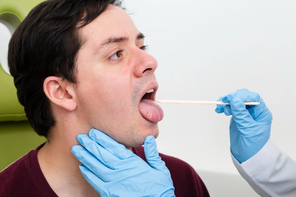
That White Patch in Your Mouth Could Be More Serious Than You Think
Most people notice a white patch inside their mouth and assume it’s a harmless canker sore or minor irritation. But when that patch doesn’t go away within two weeks — it could be leukoplakia, a potentially serious oral condition that demands medical attention. Understanding leukoplakia causes, symptoms, and treatment could literally save your life — because in some cases, leukoplakia is the earliest visible warning sign of oral cancer.
📋 Table of Contents
- → What Is Leukoplakia?
- → Types of Leukoplakia
- → Leukoplakia Symptoms: What Does It Look Like?
- → What Causes Leukoplakia?
- → Who Is Most at Risk?
- → How Is Leukoplakia Diagnosed?
- → Can Leukoplakia Turn Into Cancer?
- → Leukoplakia Treatment Options
- → How to Prevent Leukoplakia
- → Frequently Asked Questions
What Is Leukoplakia?
Leukoplakia (pronounced loo-ko-PLAY-kee-uh) is a condition in which thick, white or grayish patches form on the inner surfaces of the mouth — most commonly on the gums, cheeks, bottom of the mouth, or tongue. The word itself comes from Greek: “leuko” meaning white, and “plakia” meaning patch.
Unlike the harmless white spots caused by accidentally biting your cheek or a mild fungal infection, leukoplakia patches cannot be scraped or rubbed off. They are caused by an abnormal thickening and overgrowth of the oral mucosa — the moist tissue lining the inside of the mouth — in response to chronic irritation or other triggers.
Leukoplakia affects approximately 1 to 2% of the global population, with rates significantly higher in tobacco users. It is most commonly diagnosed in adults over 40 — particularly men — though it can affect anyone. What makes leukoplakia medically significant is that in approximately 3 to 17.5% of cases, it undergoes malignant transformation into oral squamous cell carcinoma — the most common form of oral cancer.
Consider the experience of a 52-year-old man who had smoked for 30 years. He noticed a white patch on the inside of his cheek that had been there for three months. He assumed it was nothing. When he finally mentioned it to his dentist during a routine checkup, a biopsy revealed severe dysplasia — precancerous cellular changes — that required immediate surgical treatment. Had he waited longer, the outcome could have been far worse.
Types of Leukoplakia
Not all leukoplakia is the same — and the type significantly influences the risk of malignant transformation and the urgency of treatment.
1. Homogeneous Leukoplakia
The most common and least concerning type. Appears as uniformly white, flat patches with a consistent texture and well-defined borders. The surface may be smooth, wrinkled, or finely ridged. Homogeneous leukoplakia carries a relatively lower risk of malignant transformation — but it still requires monitoring and investigation.
2. Non-Homogeneous Leukoplakia
A more concerning type with irregular texture and mixed appearance. Non-homogeneous leukoplakia includes several subtypes:
- Erythroleukoplakia (speckled leukoplakia) — Mixed red and white patches; carries a significantly higher malignant transformation risk than purely white patches
- Nodular leukoplakia — White rounded projections on a red or white base
- Verrucous leukoplakia — Thick, warty, raised white patches with an irregular surface; particularly high risk
3. Proliferative Verrucous Leukoplakia (PVL)
A rare but aggressive form of leukoplakia that tends to spread across multiple areas of the mouth simultaneously. PVL has a very high rate of malignant transformation — estimated at 60 to 70% over time — and is notoriously difficult to treat due to its tendency to recur after removal. It is more common in older women and those without traditional risk factors like tobacco use.
4. Hairy Leukoplakia
A distinct type caused by the Epstein-Barr virus (EBV) — almost exclusively seen in immunocompromised individuals, particularly those with HIV/AIDS. It appears as white, fuzzy or “hairy”-looking patches typically on the sides of the tongue. Unlike other forms, hairy leukoplakia is not considered premalignant — but its presence is a significant indicator of immune system compromise requiring medical evaluation.
| Type | Appearance | Cancer Risk |
|---|---|---|
| Homogeneous | Uniform white, flat, smooth or finely ridged | Lower (but still present) |
| Erythroleukoplakia | Mixed red and white patches | High |
| Verrucous | Thick, warty, raised white surface | High |
| Proliferative Verrucous (PVL) | Multiple spreading white warty patches | Very High (60–70%) |
| Hairy Leukoplakia | White fuzzy patches on tongue sides | Not premalignant — indicates immunosuppression |
Leukoplakia Symptoms: What Does It Look Like?
Leukoplakia itself is often asymptomatic in its early stages — meaning many people have it without experiencing any discomfort. This is precisely why it is frequently discovered incidentally during routine dental examinations rather than by the patient seeking help for symptoms.
Visual Characteristics
- White or grayish-white patches — The defining feature; appearing on the gums, inner cheeks, floor of the mouth, tongue, or roof of the mouth
- Cannot be wiped away — Unlike oral thrush (fungal infection), leukoplakia patches do not scrape off with a tongue depressor or finger
- Irregular or well-defined borders — Depending on the type; some patches have clearly defined edges, others blend gradually into surrounding tissue
- Variable texture — Flat and smooth in homogeneous type; thick, raised, or warty in non-homogeneous types
- Variable size — From a few millimeters to several centimeters; can be single or multiple patches
- Red areas within the patch — The presence of red areas (erythroplakia) mixed with the white patches significantly elevates concern for dysplasia or malignancy
Symptoms That May Accompany Leukoplakia
- Burning sensation — Particularly when eating spicy, acidic, or hot foods
- Sensitivity — The affected area may feel tender to touch or temperature
- Stiffness or restricted mouth opening — In advanced or widespread cases, particularly submucous fibrosis associated with betel nut chewing
- Numbness — A concerning symptom that may indicate deeper tissue involvement or malignant transformation
⚠️ See a Doctor or Dentist Immediately If:
- A white or red patch in your mouth does not resolve within 2 weeks
- The patch is growing in size or changing in appearance
- You notice hardening, thickening, or ulceration within the patch
- You experience unexplained bleeding from the oral tissue
- You develop difficulty swallowing or speaking
- Any patch is accompanied by numbness or pain that doesn’t resolve
What Causes Leukoplakia?
The exact mechanism of leukoplakia development involves chronic irritation or injury to the oral mucosa that triggers an abnormal protective thickening response in the epithelial cells lining the mouth. Over time, this thickening can undergo cellular changes that, in some cases, progress toward malignancy.
1. Tobacco — The Primary Cause
Tobacco use in all forms is the single most significant cause of leukoplakia. Cigarette smoking, cigar smoking, pipe smoking, chewing tobacco, and smokeless tobacco products all cause chronic chemical and thermal irritation to the oral mucosa. Studies show that tobacco users are 6 times more likely to develop leukoplakia than non-users. The risk is dose-dependent — the longer and heavier the tobacco use, the greater the risk. Importantly, leukoplakia often regresses when tobacco use is stopped — confirming the causal relationship.
2. Alcohol
Chronic alcohol consumption is an independent risk factor for leukoplakia — and its combination with tobacco creates a synergistic effect that dramatically multiplies the risk of both leukoplakia and oral cancer. Alcohol acts as a solvent, increasing the penetration of tobacco carcinogens into oral mucosal cells and directly damaging cellular DNA.
3. Betel Nut (Areca Nut) Chewing
Betel nut chewing — extremely common in South and Southeast Asia, parts of Africa, and Pacific island communities — is a major cause of leukoplakia in these populations. The combination of areca nut, betel leaf, and slaked lime produces compounds that cause chronic mucosal irritation and a particularly aggressive form of leukoplakia. It is also the primary cause of oral submucous fibrosis — a related precancerous condition causing progressive mouth stiffness.
4. Chronic Mechanical Irritation
Persistent physical trauma to the oral mucosa can trigger leukoplakia in some cases:
- Ill-fitting dentures that chronically rub against the gum or cheek tissue
- Sharp or broken teeth causing repeated injury to the tongue or cheek
- Chronic cheek biting habits
- Rough dental restorations with sharp edges
5. Human Papillomavirus (HPV)
Certain strains of HPV — particularly HPV-16 and HPV-18 — are increasingly recognized as contributing factors in some cases of oral leukoplakia and oral cancer. HPV-related oral lesions are rising significantly in incidence, particularly in non-smokers, and represent a growing public health concern.
6. Idiopathic Leukoplakia
In approximately 20 to 30% of cases, no identifiable cause can be found — this is termed idiopathic leukoplakia. Paradoxically, idiopathic leukoplakia carries a higher risk of malignant transformation than tobacco-associated leukoplakia — possibly because the driving mechanism is more deeply embedded in cellular biology rather than an external, removable irritant.
Who Is Most at Risk?
| Risk Factor | Why It Increases Risk |
|---|---|
| Tobacco use (any form) | Primary cause; 6x increased risk; dose-dependent |
| Heavy alcohol consumption | Direct mucosal damage; synergistic with tobacco |
| Betel nut chewing | Major cause in South/Southeast Asian populations |
| Male gender | Men are diagnosed 3x more often than women |
| Age over 40 | Cumulative exposure to risk factors over time |
| HIV/AIDS or immunosuppression | Significantly increases hairy leukoplakia risk |
| HPV infection | Particularly HPV-16 and HPV-18 strains |
| Poor oral hygiene | Creates environment for chronic mucosal irritation |
💡 Did You Know?
Leukoplakia patches located on the floor of the mouth, the ventral (underside) surface of the tongue, and the soft palate carry a significantly higher risk of malignant transformation than patches in other oral locations. This is because these areas have thinner, more permeable mucosal tissue — making them more vulnerable to carcinogen penetration and cellular damage.
How Is Leukoplakia Diagnosed?
Diagnosing leukoplakia requires more than simply looking at a white patch and calling it by name. A thorough diagnostic process is essential — both to confirm the diagnosis and, critically, to assess the degree of cellular abnormality (dysplasia) present, which determines the risk of malignant transformation and guides treatment decisions.
Clinical Examination
A dentist or oral medicine specialist will conduct a thorough examination of the entire oral cavity — including all mucosal surfaces, the tongue, floor of the mouth, and throat. The location, size, color, texture, and border characteristics of any suspicious patches are carefully documented. A patch that has been present for more than 2 weeks and cannot be attributed to an obvious cause warrants further investigation.
Elimination of Potential Causes
Before biopsy, clinicians typically attempt to identify and eliminate potential causes of the white patch — adjusting ill-fitting dentures, smoothing sharp teeth, stopping tobacco use — and re-examine after 2 to 4 weeks. If the patch resolves, it was likely a reactive lesion rather than true leukoplakia. If it persists, biopsy is mandatory.
Biopsy — The Gold Standard
A tissue biopsy is the definitive diagnostic test for leukoplakia. A small sample of the suspicious tissue is taken — under local anaesthesia — and examined by a pathologist under a microscope. The pathologist assesses the degree of epithelial dysplasia — the extent of abnormal cellular changes — which is classified as:
- No dysplasia — Benign; low malignant transformation risk
- Mild dysplasia — Abnormal cells in the lower third of the epithelium; regular monitoring required
- Moderate dysplasia — Abnormal cells in the lower two-thirds; higher risk; treatment usually recommended
- Severe dysplasia / Carcinoma in situ — Abnormal cells throughout the full thickness; very high malignant transformation risk; immediate treatment essential
Additional Investigations
- Toluidine blue vital staining — A dye that selectively stains dysplastic and malignant tissue more intensely than normal tissue; used as a chairside screening tool to identify areas most suspicious for dysplasia before biopsy
- Brush cytology — Non-invasive collection of cells from the lesion surface using a brush; useful for initial screening but cannot replace formal biopsy
- Fluorescence visualization — Special light devices that cause abnormal oral tissue to lose its natural fluorescence — helping identify suspicious areas not visible under white light
Can Leukoplakia Turn Into Cancer?
This is the question that makes leukoplakia a serious medical concern rather than a minor nuisance — and the honest answer is: yes, it can. Leukoplakia is classified as a potentially malignant oral disorder (PMOD) — meaning it has a measurable risk of transforming into oral squamous cell carcinoma over time.
The overall malignant transformation rate of leukoplakia is estimated at 3 to 17.5% over the lifetime of the lesion — but this figure varies enormously depending on the type of leukoplakia, the degree of dysplasia present, the location, and the patient’s ongoing risk factor exposure.
| Feature | Higher Cancer Risk | Lower Cancer Risk |
|---|---|---|
| Appearance | Non-homogeneous, red-and-white (erythroleukoplakia) | Homogeneous white only |
| Location | Floor of mouth, tongue underside, soft palate | Buccal mucosa (cheeks), hard palate |
| Dysplasia grade | Moderate to severe | None or mild |
| Cause | Idiopathic (no known cause) | Tobacco-related (may regress with cessation) |
| Patient gender | Female patients (relatively) | Male patients |
| Type | Proliferative verrucous leukoplakia | Homogeneous single patch |
Leukoplakia Treatment Options
The management of leukoplakia is tailored to the individual based on the type of lesion, degree of dysplasia present, location, size, and the patient’s overall risk profile. There is currently no universally curative treatment for leukoplakia — and high recurrence rates after treatment remain a significant clinical challenge.
1. Risk Factor Elimination — The Most Important First Step
For tobacco-associated leukoplakia, complete cessation of tobacco use is the single most important intervention. Studies show that leukoplakia regresses or resolves completely in a significant proportion of patients who stop tobacco use — with the greatest regression seen in smaller, homogeneous lesions without dysplasia. Similarly, stopping betel nut chewing, limiting alcohol intake, and eliminating mechanical irritation sources are essential first steps for all patients.
2. Surgical Excision
Surgical removal of leukoplakia patches — particularly those with moderate to severe dysplasia — is a widely used treatment approach. Options include:
- Conventional scalpel excision — Traditional surgical removal under local anaesthesia; allows complete histopathological examination of the removed tissue
- CO2 laser ablation — The most commonly used technique for leukoplakia treatment; offers precise removal with minimal bleeding, reduced postoperative pain, and excellent wound healing. Particularly valuable for large or multiple lesions.
- Electrosurgery and cryotherapy — Less commonly used alternatives; cryotherapy uses extreme cold to destroy abnormal tissue
An important caveat: surgical removal does not guarantee cure. Recurrence rates after excision range from 10 to 35%, and new lesions can develop in adjacent areas — particularly in patients who continue tobacco use or have underlying genetic susceptibility. Lifelong surveillance is essential after any leukoplakia treatment.
3. Medical Treatment
- Topical or systemic retinoids (Vitamin A derivatives) — Have shown some efficacy in promoting regression of leukoplakia by normalizing epithelial cell differentiation. However, side effects and high recurrence rates after stopping therapy limit their widespread use.
- Antifungal treatment — When Candida infection is identified as a co-existing factor (candidal leukoplakia), antifungal therapy with fluconazole or nystatin can reduce lesion severity.
- Photodynamic therapy (PDT) — An emerging treatment in which a photosensitizing agent is applied to the lesion and then activated by a specific wavelength of light, selectively destroying dysplastic cells. Promising early results with low scarring risk.
- Antiviral therapy — For hairy leukoplakia in immunocompromised patients, antiviral medications targeting EBV (acyclovir, valacyclovir) can cause temporary regression.
4. Active Surveillance (Watch and Wait)
For small, homogeneous lesions without dysplasia in low-risk locations, a carefully structured monitoring program — with clinical review every 3 to 6 months — may be appropriate as an initial approach, particularly after risk factor elimination. Any change in size, appearance, or symptoms should prompt immediate re-biopsy.
How to Prevent Leukoplakia
Given that the vast majority of leukoplakia cases are directly attributable to avoidable risk factors — particularly tobacco and alcohol — prevention is highly achievable for most people. These are not complex interventions — they are lifestyle choices with profound consequences for oral and overall health.
- Never use tobacco in any form — Cigarettes, cigars, pipes, chewing tobacco, snuff, and e-cigarettes all carry oral health risks. If you currently use tobacco, quitting is the single most impactful health decision you can make for your oral — and overall — health.
- Limit alcohol consumption — Follow recommended safe drinking guidelines. If you use both tobacco and alcohol, prioritize eliminating both together — the combined risk is far greater than either alone.
- Avoid betel nut products — Including paan, gutka, and similar preparations; all are strongly associated with oral leukoplakia and submucous fibrosis.
- Maintain excellent oral hygiene — Regular brushing, flossing, and professional dental cleanings reduce chronic irritation and mucosal inflammation.
- Ensure well-fitting dental appliances — Have dentures checked and relined regularly; sharp teeth or restorations should be smoothed promptly.
- Eat a diet rich in antioxidants — Vitamins A, C, and E from fruits and vegetables support healthy oral mucosal cell turnover and repair.
- Regular dental check-ups — Professional oral examinations every 6 months allow early detection of any suspicious lesions before they progress.
- HPV vaccination — HPV vaccination in eligible age groups reduces the risk of HPV-related oral lesions and cancers.
FAQ
How is leukoplakia different from oral thrush?
The key distinguishing feature is simple: oral thrush (candidiasis) can be wiped away — leukoplakia cannot. Oral thrush produces creamy white, cottage-cheese-like plaques that scrape off easily with a tongue depressor, revealing a red, sometimes bleeding surface beneath. Leukoplakia patches are firmly adherent to the underlying tissue and cannot be removed by rubbing or scraping. Additionally, oral thrush is caused by a fungal infection and typically resolves with antifungal treatment, while leukoplakia is a tissue change that requires different management.
Will leukoplakia go away on its own?
It depends on the cause. Tobacco-associated leukoplakia frequently regresses or resolves completely after tobacco cessation — particularly smaller, homogeneous lesions without significant dysplasia. However, idiopathic leukoplakia and lesions with established dysplasia are unlikely to resolve without active treatment. Never assume a white patch will resolve on its own without professional evaluation — waiting risks missing an early malignancy.
How often should leukoplakia be monitored after treatment?
After treatment — whether surgical removal or active surveillance — leukoplakia requires lifelong monitoring. Most clinicians recommend follow-up appointments every 3 months for the first year after treatment, then every 6 months indefinitely thereafter. Any change in the treated area or the appearance of new lesions elsewhere in the mouth should prompt immediate re-evaluation and possible re-biopsy. The recurrence rate of leukoplakia after treatment is significant — making ongoing vigilance non-negotiable.
Is leukoplakia painful?
In most cases, particularly early-stage homogeneous leukoplakia, the patches are completely painless — which is one of the reasons they are often ignored. Some patients experience a mild burning sensation when eating spicy or acidic foods. The development of pain, tenderness, or numbness within a leukoplakia lesion is a significant warning sign that may indicate dysplastic changes or malignant transformation — and should prompt immediate medical evaluation.
Can non-smokers get leukoplakia?
Yes — while tobacco use is the most common cause, leukoplakia can develop in non-smokers due to other factors including alcohol use, betel nut chewing, chronic mechanical irritation, HPV infection, or without any identifiable cause (idiopathic leukoplakia). Idiopathic leukoplakia in non-smokers actually carries a comparatively higher risk of malignant transformation — making it particularly important for non-smokers not to dismiss an unexplained white oral patch as harmless.
What foods should I avoid with leukoplakia?
While no specific diet treats leukoplakia, certain foods and habits can worsen mucosal irritation and should be minimized: very spicy foods, highly acidic foods and drinks, extremely hot foods and beverages, alcohol, and rough or hard-textured foods that may traumatize the affected area. Conversely, a diet rich in antioxidant vitamins (A, C, E), lycopene from tomatoes, and green tea polyphenols may support oral mucosal health — though dietary changes alone are not a substitute for medical treatment.
A White Patch Is Not Worth Ignoring — Ever
Leukoplakia sits at a uniquely important crossroads in medicine — it is a visible, accessible, early warning system that the body provides. Unlike many cancers that develop silently in organs we cannot see, leukoplakia appears in your mouth — where you, your dentist, and your doctor can see it, assess it, and act on it before it becomes something far more serious.
Understanding leukoplakia causes, symptoms, and treatment means you are equipped to take that warning seriously. Stop tobacco use. See your dentist regularly. Report any white patch that doesn’t resolve in two weeks. Get it biopsied if advised.
Your mouth is trying to tell you something. It deserves your full attention — right now, before the message becomes impossible to ignore.
Medical Disclaimer: The information provided in this article is for general educational purposes only and does not constitute medical advice, diagnosis, or treatment. Leukoplakia is a potentially serious oral condition that requires professional evaluation by a qualified dentist, oral medicine specialist, or otolaryngologist. Any persistent white patch in the mouth lasting more than two weeks should be evaluated by a healthcare professional promptly. BestInMeds.com does not endorse any specific treatment protocol, surgical technique, or healthcare provider. Always follow the guidance of your qualified medical team regarding diagnosis and management.