
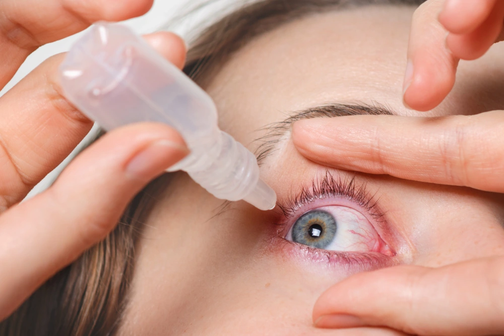
Glaucoma Is Stealing Vision Silently Millions Don’t Know They Have It
It has been called the “silent thief of sight” — and for good reason. Glaucoma is a group of eye diseases that progressively damage the optic nerve, causing irreversible vision loss that often goes completely unnoticed until a significant portion of sight is already gone forever. With over 80 million people affected worldwide, understanding glaucoma symptoms, causes, and treatment is not just important for the elderly — it is essential health knowledge for everyone.
📋 Table of Contents
- → What Is Glaucoma?
- → Types of Glaucoma You Must Know
- → What Causes Glaucoma?
- → Glaucoma Symptoms: What to Watch For
- → Who Is Most at Risk?
- → How Is Glaucoma Diagnosed?
- → Complications and Long-Term Impact
- → Glaucoma Treatment Options
- → How to Protect Your Eyes from Glaucoma
- → Frequently Asked Questions
What Is Glaucoma?
Glaucoma is not a single disease — it is a group of related eye conditions that share one defining feature: progressive damage to the optic nerve, the critical structure that transmits visual information from the eye to the brain. When the optic nerve is damaged, the brain stops receiving complete visual signals — and vision is lost, beginning with peripheral (side) vision and potentially progressing to total blindness.
What makes glaucoma particularly dangerous is its characteristic silence. In the most common form — open-angle glaucoma — there are no pain signals, no redness, no obvious warning signs. Vision loss occurs so gradually that the brain compensates, and patients often don’t notice significant vision impairment until 40% or more of their optic nerve fibers have already been permanently destroyed.
According to the World Health Organization, glaucoma is the second leading cause of blindness worldwide — responsible for approximately 8% of all global blindness cases. More alarming still, the WHO estimates that 50% of people with glaucoma are unaware they have it. This is the public health challenge at the heart of glaucoma awareness — a disease that is treatable when caught early but devastating when missed.
Types of Glaucoma You Must Know
Understanding the different types of glaucoma is essential because each type has distinct characteristics, symptoms, and treatment approaches. Receiving the correct diagnosis is the foundation of effective management.
1. Primary Open-Angle Glaucoma (POAG)
This is by far the most common form — accounting for approximately 70–90% of all glaucoma cases. In POAG, the drainage angle of the eye (the structure through which fluid exits) remains open and appears structurally normal, but drainage efficiency gradually decreases over time. This causes a slow, painless buildup of intraocular pressure (IOP) that silently damages the optic nerve over years or decades. Most people have absolutely no symptoms until significant vision loss has already occurred.
2. Acute Angle-Closure Glaucoma
Unlike open-angle glaucoma, angle-closure glaucoma is a dramatic medical emergency. It occurs when the drainage angle between the iris and cornea closes suddenly, causing a rapid and extreme spike in intraocular pressure. Symptoms appear abruptly and severely — intense eye pain, headache, nausea, vomiting, blurred vision, and seeing halos around lights. Without emergency treatment within hours, permanent vision loss can occur. This type is more common in people of Asian descent and those with naturally narrow anterior chambers.
3. Normal-Tension Glaucoma (NTG)
One of the most puzzling forms of glaucoma — NTG causes optic nerve damage despite intraocular pressure remaining within the statistically normal range (below 21 mmHg). It is believed that in NTG, the optic nerve is abnormally sensitive to even normal pressure levels, or that poor blood flow to the optic nerve is the primary driver of damage. NTG is particularly prevalent in people of Japanese descent and women.
4. Secondary Glaucoma
Secondary glaucoma develops as a consequence of another eye condition or systemic disease — including eye trauma, uveitis (eye inflammation), diabetic retinopathy, prolonged corticosteroid use, or advanced cataracts. Treatment of secondary glaucoma requires addressing both the elevated pressure and the underlying condition driving it.
5. Congenital Glaucoma
A rare but serious form present from birth — caused by abnormal development of the eye’s drainage system during fetal development. Symptoms in infants include excessive tearing, light sensitivity, enlarged and cloudy eyes, and frequent eye rubbing. Early surgical intervention is essential to prevent permanent vision impairment.
| Type | Onset | Symptoms | Emergency? |
|---|---|---|---|
| Open-Angle | Gradual — years | None until late stage | No — but urgent monitoring needed |
| Angle-Closure | Sudden — hours | Severe pain, nausea, halos | YES — immediate care |
| Normal-Tension | Gradual — years | None until late stage | No — but requires close monitoring |
| Secondary | Varies | Depends on cause | Sometimes |
| Congenital | Birth / infancy | Tearing, light sensitivity, cloudy eyes | Yes — early surgery critical |
What Causes Glaucoma?
The fundamental mechanism of most glaucoma involves elevated intraocular pressure (IOP) — the fluid pressure inside the eye. To understand why this pressure rises, it helps to understand how normal eye fluid circulation works.
The eye continuously produces a clear fluid called aqueous humor in the ciliary body. This fluid flows through the pupil into the anterior chamber and drains out through the trabecular meshwork — a sponge-like drainage structure at the angle where the iris meets the cornea. When this drainage system becomes partially blocked or inefficient, fluid accumulates and pressure builds — like a sink with a partially blocked drain.
Elevated IOP compresses and damages the delicate nerve fibers of the optic nerve — particularly at the optic nerve head (the point where the nerve exits the eye). Once these nerve fibers die, they cannot regenerate. The damage is permanent and cumulative.
Key Contributing Factors
- Reduced trabecular meshwork drainage efficiency — The primary cause of open-angle glaucoma; the drainage angle becomes progressively less efficient with age
- Anatomical predisposition — A naturally narrow anterior chamber angle predisposes to angle-closure glaucoma
- Genetics — Multiple gene variants associated with elevated IOP and optic nerve vulnerability have been identified; first-degree relatives of glaucoma patients have a 4 to 9 times higher risk
- Corticosteroid use — Long-term use of steroid eye drops, oral steroids, or even nasal or inhaled steroids can elevate IOP and trigger steroid-induced glaucoma
- Eye trauma — Physical injury to the eye can damage the drainage structures, leading to traumatic glaucoma years later
- Systemic diseases — Diabetes, hypertension, and cardiovascular disease are all associated with increased glaucoma risk through their effects on ocular blood flow and nerve health
- Vascular dysregulation — Poor blood flow to the optic nerve — particularly in normal-tension glaucoma — is an increasingly recognized cause of nerve damage independent of pressure
Glaucoma Symptoms: What to Watch For
This is where glaucoma becomes particularly treacherous. The most common form — open-angle glaucoma — produces no symptoms whatsoever in its early and middle stages. By the time a patient notices something is wrong with their vision, irreversible damage has already occurred. This is precisely why regular eye examinations are so critically important — especially for those in high-risk groups.
Late-Stage Open-Angle Glaucoma Symptoms
- Peripheral vision loss — The earliest detectable symptom; blind spots gradually develop in the side vision, often going unnoticed because the brain fills in the gaps
- Tunnel vision — As the disease progresses, the visual field narrows progressively until only a small central island of vision remains
- Difficulty with contrast and low-light vision — Reduced ability to see in dim light or distinguish subtle contrasts
- Frequent prescription changes — Needing increasingly stronger glasses that still don’t seem to help clearly
Acute Angle-Closure Glaucoma — Emergency Symptoms
⚠️ Go to Emergency Immediately If You Experience:
- Sudden, severe eye pain — Often described as the worst eye pain ever experienced
- Intense headache on one side — accompanying the eye pain
- Halos around lights — Rainbow-colored rings appearing around light sources
- Sudden blurred or hazy vision in the affected eye
- Nausea and vomiting accompanying eye pain — often mistaken for migraine or gastrointestinal illness
- Redness of the eye — Often with a mid-dilated, fixed pupil
Acute angle-closure glaucoma is one of medicine’s true emergencies. Every hour of delay in treatment increases the risk of permanent, irreversible vision loss. If you or anyone around you experiences these symptoms — go to the nearest emergency eye clinic immediately.
Who Is Most at Risk?
While glaucoma can develop in anyone, certain characteristics dramatically increase the likelihood. Knowing your risk profile is the most empowering step you can take — because it determines how frequently you need eye examinations and when preventive monitoring should begin.
| Risk Factor | Level of Risk Increase |
|---|---|
| Age over 60 | Risk increases 6-fold after age 60 |
| Family history of glaucoma | 4–9 times higher risk than general population |
| African or Hispanic descent | 3–4 times higher risk; earlier onset |
| Asian descent | Higher risk of angle-closure type specifically |
| High intraocular pressure (ocular hypertension) | Primary modifiable risk factor |
| Diabetes mellitus | 2 times higher risk |
| High myopia (nearsightedness) | Significantly increased open-angle risk |
| Long-term corticosteroid use | Can cause steroid-induced IOP elevation |
| Previous eye injury or surgery | Traumatic glaucoma can develop years later |
| Thin central cornea | Associated with higher glaucoma risk and may cause IOP underestimation |
💡 Important Age Milestone
The American Academy of Ophthalmology recommends that all adults have a comprehensive baseline eye examination by age 40 — even without symptoms or known risk factors. For African Americans and those with a family history of glaucoma, this baseline exam should occur by age 35. After 65, annual eye exams are strongly recommended for everyone. Early detection is the only reliable way to prevent glaucoma-related blindness.
How Is Glaucoma Diagnosed?
Because glaucoma produces no early symptoms, diagnosis depends entirely on proactive eye examinations. A comprehensive glaucoma evaluation involves multiple tests — no single test is sufficient to diagnose or rule out glaucoma with confidence.
Diagnostic Tests Used
- Tonometry (IOP measurement) — Measures the pressure inside the eye using an air puff or contact tonometer. Normal IOP is generally considered 10–21 mmHg, though glaucoma can occur at any pressure level.
- Ophthalmoscopy (optic nerve examination) — Direct examination of the optic nerve head through a dilated pupil. The doctor looks for characteristic changes — increased cup-to-disc ratio, rim thinning, hemorrhages, and asymmetry between eyes.
- Visual field testing (perimetry) — Maps the patient’s full visual field to detect characteristic glaucomatous blind spots and peripheral vision loss. Automated perimetry is the standard clinical tool.
- Optical Coherence Tomography (OCT) — Advanced imaging technology that measures the thickness of the retinal nerve fiber layer with extraordinary precision. Can detect glaucomatous thinning years before visual field loss becomes apparent — making it invaluable for early diagnosis.
- Gonioscopy — Examines the drainage angle of the eye directly using a special contact lens. Essential for determining whether glaucoma is open-angle or angle-closure type.
- Pachymetry (corneal thickness measurement) — Measures central corneal thickness; important because thin corneas are associated with higher glaucoma risk and can cause IOP to be underestimated by tonometry.
Complications and Long-Term Impact
The most devastating complication of untreated or inadequately managed glaucoma is permanent, irreversible blindness. Because optic nerve fibers cannot regenerate once destroyed, every stage of vision loss from glaucoma is permanent. This is the defining tragedy of this disease — and the primary driver of the urgent need for early detection and consistent treatment.
- Progressive peripheral vision loss — Gradually narrowing visual field that, when severe, leaves only a small tunnel of central vision
- Total blindness — End-stage glaucoma causes complete loss of all functional vision — a fate that is entirely preventable with early diagnosis and treatment
- Impact on daily function — Even moderate glaucomatous vision loss significantly impairs driving ability, reading, mobility, and independence — dramatically reducing quality of life
- Falls and accidents — Peripheral vision loss from glaucoma is a major risk factor for falls and road traffic accidents in older adults
- Psychological impact — Depression, anxiety, and social isolation are significantly more common in people living with glaucoma — particularly as vision loss progresses
- Treatment side effects — Long-term glaucoma medications and surgical interventions carry their own complication risks that require ongoing management
Glaucoma Treatment Options
The fundamental goal of all glaucoma treatment is singular and uncompromising: lower intraocular pressure to a level that halts or slows optic nerve damage. While lost vision cannot be restored, treatment can effectively prevent further deterioration — preserving the vision that remains. The sooner treatment begins, the more vision is protected.
1. Eye Drops — First-Line Treatment
Medicated eye drops are the most common initial treatment for open-angle glaucoma. They work by either reducing the production of aqueous humor or increasing its drainage from the eye — both of which lower IOP effectively.
- Prostaglandin analogs (latanoprost, bimatoprost, travoprost) — The most commonly prescribed first-line drops; increase aqueous outflow. Applied once daily at bedtime. Side effects include darkening of the iris and eyelashes.
- Beta-blockers (timolol, betaxolol) — Reduce aqueous production. Used twice daily. Contraindicated in asthma and certain heart conditions.
- Alpha-2 agonists (brimonidine) — Both reduce production and increase drainage. Also have a neuroprotective effect on the optic nerve.
- Carbonic anhydrase inhibitors (dorzolamide, brinzolamide) — Reduce aqueous production. Available as eye drops and oral tablets.
- Rho kinase inhibitors (netarsudil) — A newer class that increases trabecular outflow; particularly effective in normal-tension glaucoma.
Adherence to eye drop therapy is one of the greatest challenges in glaucoma management. Studies show that up to 50% of glaucoma patients do not use their drops as prescribed — and poor adherence is the leading cause of preventable vision loss in treated patients.
2. Laser Treatment
- Selective Laser Trabeculoplasty (SLT) — Uses targeted laser energy to improve trabecular meshwork drainage. Highly effective with minimal side effects; increasingly recommended as a first-line alternative to eye drops. Effects typically last 3–5 years and the procedure can be repeated.
- Laser Peripheral Iridotomy (LPI) — Creates a small opening in the peripheral iris to allow fluid to bypass a narrow drainage angle — the definitive preventive treatment for angle-closure glaucoma.
- Cyclophotocoagulation — Laser treatment targeting the ciliary body to reduce aqueous production; used in advanced glaucoma cases where other treatments have failed.
3. Surgical Treatment
- Trabeculectomy — The traditional gold standard glaucoma surgery. Creates a new drainage pathway (bleb) under the conjunctiva, allowing fluid to bypass the blocked trabecular meshwork entirely. Very effective at lowering IOP but requires close postoperative monitoring.
- Glaucoma Drainage Devices (tube shunts) — Small silicone tubes implanted to redirect aqueous humor to an external reservoir plate; used when trabeculectomy has failed or is likely to fail.
- Minimally Invasive Glaucoma Surgery (MIGS) — A newer category of micro-scale surgical procedures that lower IOP with significantly less risk and faster recovery than traditional surgery. Includes trabecular micro-bypass stents (iStent), canaloplasty, and goniotomy. Often performed simultaneously with cataract surgery.
How to Protect Your Eyes from Glaucoma
While the genetic component of glaucoma cannot be changed, there are meaningful steps everyone can take to reduce their risk and ensure the earliest possible detection if the disease does develop.
Regular Comprehensive Eye Examinations
This is the single most important preventive action. Regular eye examinations — including IOP measurement, optic nerve assessment, and visual field testing — are the only reliable way to detect glaucoma before significant vision loss occurs. Frequency recommendations vary by age and risk profile, but most people over 40 should have a comprehensive eye exam at least every 2 years, and annually after 60.
Lifestyle Modifications That Support Eye Health
- Regular aerobic exercise — Multiple studies confirm that regular moderate exercise (brisk walking, cycling, swimming) reduces IOP by 20–25% and improves ocular blood flow. Even 30 minutes of aerobic exercise three times per week produces measurable IOP-lowering effects.
- Healthy diet rich in antioxidants — Vitamins C, E, and A, omega-3 fatty acids, and lutein/zeaxanthin support optic nerve health and reduce oxidative damage to retinal cells. Dark leafy greens, colorful vegetables, fatty fish, and nuts are particularly beneficial.
- Avoid extreme inversion exercises — Headstands, yoga inversions, and certain weightlifting techniques dramatically elevate IOP temporarily — a concern for those already at risk.
- Manage systemic health conditions — Controlling blood pressure, blood sugar, and maintaining cardiovascular health all support healthy ocular blood flow and reduce glaucoma risk.
- Avoid prolonged steroid use without monitoring — If long-term corticosteroid therapy is medically necessary, regular IOP monitoring by an ophthalmologist is essential.
- Protect eyes from injury — Wear appropriate eye protection during sports and high-risk work activities to prevent traumatic glaucoma.
- Know your family history — Inform your ophthalmologist if any first-degree relative has been diagnosed with glaucoma — this changes your monitoring schedule significantly.
Frequently Asked Questions
Can glaucoma be cured completely?
Currently, there is no cure for glaucoma — and vision already lost to the disease cannot be restored. However, glaucoma is very effectively managed with treatment. With appropriate and consistent therapy — whether eye drops, laser, or surgery — the vast majority of glaucoma patients retain useful vision for their entire lifetime. The key is early detection and unwavering adherence to treatment. Think of glaucoma management like blood pressure management — it requires lifelong attention but is very successfully controlled.
Does high eye pressure always mean glaucoma?
No — and this is an important distinction. Elevated IOP (ocular hypertension) does not automatically mean glaucoma is present. Some people have persistently elevated pressure without any optic nerve damage — a condition called ocular hypertension. Conversely, glaucoma can occur with normal IOP (normal-tension glaucoma). Diagnosis requires assessment of the optic nerve, visual field, and multiple other parameters — not IOP measurement alone. However, ocular hypertension does significantly increase the risk of developing glaucoma and warrants regular monitoring.
Is glaucoma hereditary?
Yes — genetics plays a significant role in glaucoma risk. Having a first-degree relative (parent, sibling) with glaucoma increases your risk by 4 to 9 times compared to the general population. Multiple gene variants associated with elevated IOP, optic nerve vulnerability, and drainage angle anatomy have been identified. If glaucoma runs in your family, inform your ophthalmologist and begin regular eye examinations earlier than the general population recommendations — ideally by age 35.
Can glaucoma affect young people?
Yes — while glaucoma is far more common in older adults, it can affect people of any age. Juvenile open-angle glaucoma is a genetic form that develops in teenagers and young adults. Congenital glaucoma affects newborns. Additionally, secondary glaucoma from eye trauma, steroid use, or inflammatory conditions can develop at any age. Young people who experience unexplained vision changes, eye pain, or who have a strong family history of glaucoma should seek ophthalmological evaluation regardless of age.
How often should I have eye exams if I have glaucoma?
The frequency of monitoring for diagnosed glaucoma depends on disease severity and stability. Most glaucoma patients require eye exams every 3 to 6 months — including IOP measurement, visual field testing, and OCT imaging to monitor optic nerve fiber layer thickness. Stable, well-controlled glaucoma may be monitored every 6 months. Active disease, recent treatment changes, or elevated pressure requires more frequent visits. Never skip scheduled appointments — glaucoma can progress silently even when you feel fine.
Are glaucoma eye drops safe for long-term use?
Glaucoma eye drops are generally safe for long-term use — and for most patients, the vision-preserving benefits far outweigh the risks of side effects. However, some side effects do occur with prolonged use: prostaglandin analogs can darken the iris and cause periorbital fat loss; beta-blockers can worsen asthma and lower heart rate; preservatives in multi-dose drops (particularly benzalkonium chloride) can irritate the ocular surface over time. Preservative-free formulations are available for sensitive patients. Always discuss any side effects with your ophthalmologist — there are usually alternative options available.
Don’t Wait Until You Notice — By Then It May Be Too Late
Glaucoma’s greatest weapon is its silence. It steals your vision so gradually, so painlessly, that by the time you notice something is wrong — the damage is already done and cannot be undone. This is the heartbreaking reality of a disease that is almost always preventable in its worst consequences.
Understanding glaucoma symptoms, causes, and treatment is your first line of defense. But knowledge alone is not enough — you must act on it. Book that eye examination you’ve been putting off. Tell your ophthalmologist if glaucoma runs in your family. Take your eye drops every single day if you’ve been prescribed them. Show up for every scheduled monitoring appointment.
Your vision is irreplaceable. Glaucoma is beatable — but only if you give medicine the chance to fight it before the damage is done.
Medical Disclaimer: The information provided in this article is for general educational purposes only and does not constitute medical advice, diagnosis, or treatment. Glaucoma is a serious medical condition requiring professional evaluation and management by a qualified ophthalmologist. If you are experiencing any eye symptoms, vision changes, or have risk factors for glaucoma, schedule a comprehensive eye examination with a licensed eye care professional as soon as possible. BestInMeds.com does not endorse any specific medication, surgical technique, or eye care provider. Individual treatment plans must be tailored by a qualified healthcare professional based on each patient’s unique clinical situation.